Glycation and AGEs: How sugar silently ages your tissues
AGEs cross-link proteins, stiffen arteries, and amplify inflammation. Learn how glycation forms, how to track it, and practical ways to slow new AGE buildup.
Every time you eat a grilled steak, drink a caramel latte, or let your blood sugar spike after a meal, a slow chemical reaction is happening inside your body. Glucose molecules can attach to proteins, DNA, and lipids — forming molecular damage called advanced glycation end products (AGEs). If you want to understand precisely how glucose spikes connect to the hallmarks of aging, that guide covers the broader mechanistic picture.
Unlike many short-lived forms of cellular damage, AGE cross-links are slow to clear. They accumulate in long-lived tissues. By later adulthood, skin collagen usually contains several times more AGE signal than it did in young adulthood. Arteries stiffen. Collagen cross-links. Kidneys filter less efficiently. And the inflammation triggered by AGEs can feed into other aging pathways — from mitochondrial dysfunction to epigenetic drift.
Here’s the uncomfortable truth: glycation isn’t something that only happens to people with diabetes. It’s a universal process that tends to accelerate with age, high-heat cooking, smoking, insulin resistance, and chronic blood sugar elevations — including values that may still look “normal” on a basic lab report.
Quick answer
Glycation is a slow sugar-protein reaction that forms advanced glycation end products, or AGEs. AGEs can cross-link collagen-rich tissues, activate RAGE-driven inflammation, and track with vascular, kidney, skin, and metabolic aging. The practical goal is not to eliminate glycation; it is to reduce the rate of new AGE formation by improving glucose control, using lower-heat cooking more often, building insulin sensitivity, and monitoring HbA1c or other glycation markers over time.
Key facts
- Glycation forms AGEs when reducing sugars react with proteins, lipids, or nucleic acids.
- AGE cross-links stiffen collagen-rich tissues such as skin, arteries, kidneys, and joints.
- RAGE signaling amplifies oxidative stress and inflammatory cytokines.
- HbA1c reflects recent glycation; skin autofluorescence reflects longer-term tissue AGE load.
- Lower-heat cooking, better insulin sensitivity, and regular exercise can reduce new AGE exposure.
What you’ll learn:
- What glycation is and how AGEs form
- The Maillard reaction: inside your body and on your plate
- How AGEs drive aging at the molecular level
- The RAGE receptor: when damage signals more damage
- 8 evidence-based strategies to reduce glycation
- How to track your glycation burden
What is glycation?
Quick definition: Glycation is a non-enzymatic chemical reaction in which sugar molecules bond to proteins, lipids, or DNA — forming advanced glycation end products (AGEs) that accumulate with age and can drive tissue damage.
Glycation follows a predictable biochemical cascade. It starts when a reducing sugar — glucose, fructose, or galactose — reacts with the amino group of a protein. This initial step, called a Schiff base formation, is reversible. If blood sugar returns to normal quickly, the reaction undoes itself.
But if glucose stays elevated for hours, the Schiff base can rearrange into a more stable compound called an Amadori product. This is what glycated hemoglobin (HbA1c) actually measures — hemoglobin proteins with glucose attached after weeks of exposure.
Over months and years, Amadori products undergo further oxidation, dehydration, and cross-linking to form AGEs — irreversible molecular structures that alter the function of whatever protein they’re attached to. Collagen becomes stiff. Elastin loses its stretch. Enzymes lose their catalytic precision.
Why glycation matters for longevity
The connection between glycation and aging is more than a correlation. A landmark 2018 review in Cell Metabolism, extended by 2025 syntheses in Antioxidants and Advanced Science, documented that AGEs interact with several hallmarks of aging:
- Genomic instability: AGEs glycate DNA directly, altering gene expression patterns
- Epigenetic alterations: RAGE-mediated inflammation shifts the epigenetic landscape
- Telomere attrition: oxidative stress from AGEs shortens telomeres faster
- Mitochondrial dysfunction: glycated mitochondrial proteins impair energy production
- Loss of proteostasis: cross-linked proteins resist normal cellular recycling
In simpler terms: AGEs do not merely mark aging; they can participate in it. Recent 2025 work on “garb-aging and inflammaging” positions RAGE as a key transmitter between accumulated molecular waste and the chronic low-grade inflammation associated with biological aging.
The Maillard reaction: inside your body and on your plate
The same chemical reaction that gives bread its golden crust and steak its savory char is happening inside your blood vessels right now. It’s called the Maillard reaction, named after French chemist Louis-Camille Maillard who described it in 1912.
In cooking, the Maillard reaction requires heat — temperatures above 250°F (120°C) — to brown sugars and proteins. In your body, the same reaction occurs at 98.6°F (37°C). It just takes longer. Instead of minutes on a grill, it takes weeks and months in your bloodstream. But the end products are chemically identical.
Exogenous AGEs: what you eat matters
Research estimates that a meaningful share of circulating AGEs comes directly from food, while the rest form endogenously — inside your body — from normal metabolic processes.
The highest dietary AGE sources, measured in kilounits per serving (kU):
| Food | AGEs (kU/serving) | Cooking Method |
|---|---|---|
| Grilled chicken breast | 5,828 | Pan-fried, high heat |
| Bacon, fried | 11,905 | Fried |
| Butter | 1,736 | — |
| Roasted almonds | 1,995 | Dry-roasted |
| Steamed vegetables | 43 | Steamed |
| Raw apple | 13 | — |
The pattern is clear: animal products cooked at high temperatures generate far more AGEs than plant foods or low-temperature cooking methods. A 2010 study in the Journal of the American Dietetic Association found that switching from a typical Western diet to a low-AGE diet reduced circulating AGE markers over several months. A 2025 Dutch cohort of 718 participants extended this by showing that higher dietary AGE intake also shifts the gut microbiome, reducing beneficial bacteria that produce short-chain fatty acids — a possible second route through which cooked-food AGEs may influence inflammation.
Fructose: the silent accelerator
Not all sugars glycate at the same rate. In laboratory systems, fructose reacts with proteins several times faster than glucose. This is particularly relevant because fructose consumption has increased over the past 50 years through high-fructose corn syrup, fruit juices, and processed foods.
Unlike glucose, fructose is metabolized largely in the liver — and when intake is high, it can increase glycation intermediates such as methylglyoxal and glyoxal.
How AGEs drive aging at the molecular level
AGEs damage your tissues through two distinct mechanisms: direct structural damage and receptor-mediated inflammation.
Mechanism 1: cross-linking and stiffening
When AGEs form on collagen — the most abundant protein in your body — they can create durable chemical cross-links between collagen fibers. Imagine collagen as a suspension bridge with cables that can flex and absorb stress. AGE cross-links are like welding some of those cables together. The bridge becomes more rigid and less resilient.
This cross-linking explains some of the most visible signs of aging:
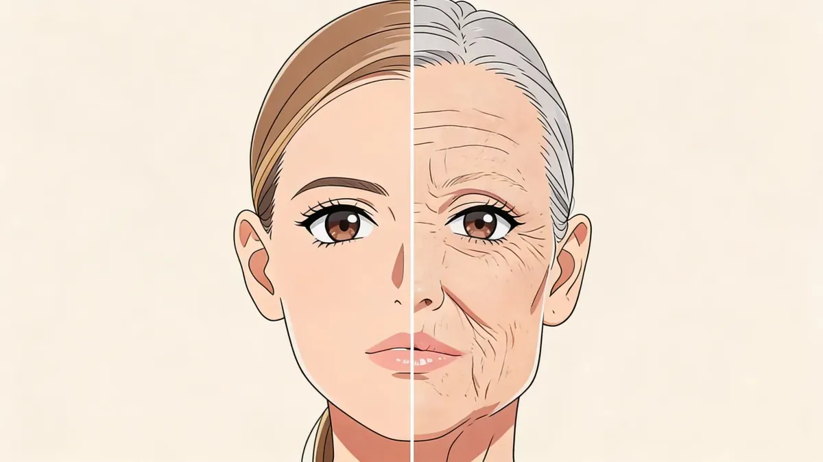
- Skin: loss of elasticity, wrinkles, yellowing — AGEs accumulate in dermal collagen and elastin, reducing skin flexibility with age. This pathway converges with UV radiation, which drives collagen degradation through matrix metalloproteinase overexpression — making glycation and sun exposure two compounding routes toward the same structural deterioration. The full science of UV-driven collagen damage explains how the two processes can amplify each other.
- Arteries: arterial stiffness — AGE cross-links in vascular collagen increase pulse wave velocity, raising systolic blood pressure and cardiovascular risk
- Kidneys: reduced filtration — glycated basement membrane proteins impair glomerular filtration, contributing to the age-related decline in kidney function
- Joints: cartilage stiffening — cross-linked cartilage collagen loses shock-absorbing capacity
The critical insight: cross-linked proteins resist degradation. Normal collagen turns over slowly. Glycated collagen can persist for years because cross-links make it harder for enzymes (MMPs) to break it down. This creates an accumulation cycle that tends to accelerate with age.
Mechanism 2: RAGE activation and chronic inflammation
The second mechanism may be even more important clinically. AGEs don’t just sit passively in your tissues — they can signal through a receptor called RAGE (Receptor for Advanced Glycation End products).
When AGEs bind to RAGE on cell surfaces, they trigger a cascade:
- NF-kB activation — the master transcription factor for inflammation
- Cytokine release — IL-6, TNF-alpha, and hs-CRP increase
- Oxidative stress — NADPH oxidase and mitochondrial pathways generate reactive oxygen species (ROS)
- More RAGE expression — inflammation upregulates RAGE itself, creating a positive feedback loop
This self-amplifying cycle — AGEs activate RAGE, which generates more oxidative stress, which creates more AGEs, which activate more RAGE — is one reason glycation damage can compound over time.
A 2021 study in Experimental & Molecular Medicine showed that blocking RAGE signaling in aged mice reduced inflammatory markers and improved age-related kidney and vascular phenotypes. The AGEs were still present — but without RAGE amplifying the signal, their impact was reduced. A 2025 review in Advanced Science went further, showing that polyphenol-rich dietary interventions may modulate RAGE expression and downstream inflammation — suggesting that what you eat may tune the receptor as much as the ligand.
Researchers are also watching the soluble decoy forms of the receptor — sRAGE and esRAGE — which circulate in blood and bind AGEs before they can reach cell-surface RAGE. Early-phase drug candidates targeting RAGE directly, including new benzo[b]thiophene-2-carboxamide antagonists reported in 2025, are now in preclinical development.
Glycation, PhenoAge, and biological aging
Several AGE-related markers feed directly into biological age calculators. Fasting glucose is a core component of the PhenoAge algorithm, and albumin — the protein most affected by glycation in blood — is the single most weighted biomarker in the Aging.ai system.
The connection is practical: higher glycation burden can reduce functional albumin and worsen the metabolic inputs that drive biological-age scores. This is why maintaining healthy blood sugar levels — not just avoiding diabetes — has such an outsized impact on biological aging calculations.
8 evidence-informed strategies to reduce glycation
AGE accumulation is inevitable, but its rate is highly modifiable. These strategies target both exogenous (dietary) and endogenous (metabolic) sources of glycation.
1. Lower your average blood sugar — even within the “normal” range
The rate of glycation rises as blood glucose exposure rises. A person with an average blood sugar of 115 mg/dL (6.4 mmol/L) generally creates more glycation pressure than someone at 85 mg/dL (4.7 mmol/L) — even though both values may fall within the “normal” clinical range.
Large cohort studies show the relationship between HbA1c and all-cause mortality is U-shaped. In people without diabetes, the lowest-risk zone often sits near the low-to-mid 5% range; levels above the prediabetes threshold raise cardiometabolic risk, while unusually low values can reflect anemia, frailty, or other confounders rather than pristine glucose control. The takeaway: aim for metabolically healthy, not artificially low.
How to do it:
- Monitor your HbA1c — discuss a healthy personal target with your clinician rather than focusing only on “under 5.7%”
- Reduce refined carbohydrates and added sugars — especially fructose-heavy sources
- Eat carbohydrates alongside protein, fat, and fiber to blunt glucose spikes
- Walk for 10-15 minutes after meals — post-meal walks reduce glucose peaks by 20-30%
- Consider a continuous glucose monitor (CGM) for 2–4 weeks — it reveals post-meal spikes invisible to fasting tests and HbA1c
Expected results: HbA1c changes usually become visible within 8-12 weeks, since it reflects the last 90 days of blood sugar exposure.
2. Change how you cook — not just what you eat
Cooking temperature and dryness are major determinants of dietary AGE content. The same chicken breast can contain far fewer AGEs when poached than when pan-fried.
How to do it:
- Prefer steaming, boiling, poaching, and stewing over grilling, frying, and roasting
- Marinate meats in acidic ingredients (lemon juice, vinegar, tomato) — this can meaningfully reduce AGE formation
- Cook at lower temperatures for longer times when possible
- Use ceramic cookware instead of bare metal surfaces
Expected results: Switching to low-AGE cooking methods can substantially reduce dietary AGE intake without changing what you eat.
3. Activate autophagy to clear glycated proteins
Autophagy is one of your body’s main mechanisms for removing damaged proteins — including glycated ones. When autophagy is active, cells can engulf and recycle AGE-modified proteins before they become part of more durable cross-linked structures.
How to do it:
- Practice time-restricted eating (14-16 hour overnight fasts)
- Include periodic 24-36 hour fasts if medically appropriate
- Exercise regularly — both resistance training and aerobic exercise activate autophagy
- Avoid constant snacking, which suppresses autophagy through chronic mTOR activation
Expected results: Autophagy activation markers can rise after prolonged overnight fasting. Regular practice may build sustained cellular cleanup capacity.
4. Improve insulin sensitivity
Insulin resistance creates a metabolic environment that accelerates glycation. When cells resist insulin’s signal, glucose remains elevated in the bloodstream for longer periods — extending the window for glycation reactions.
How to do it:
- Prioritize resistance training — muscle is your largest glucose sink
- Optimize sleep quality and duration (7-8 hours)
- Manage chronic stress (cortisol directly antagonizes insulin)
- Consider a Mediterranean dietary pattern, which consistently improves insulin sensitivity in trials
Expected results: Fasting insulin improvements can be measurable within 4-8 weeks of consistent lifestyle changes.
5. Increase dietary antioxidants and anti-glycation compounds
Certain nutrients directly interfere with AGE formation or enhance your body’s ability to detoxify glycation intermediates.
Key anti-glycation compounds:
- Carnosine (beta-alanyl-L-histidine): found in red meat and poultry, acts as a sacrificial target — glycation can attack carnosine instead of structural proteins. A 2025 skin-focused clinical study using supramolecular carnosine reported improved pigmentation-related measures over 28 days, but it should not be treated as proof of systemic anti-aging effects
- Benfotiamine (fat-soluble vitamin B1): redirects glucose away from glycation pathways and into safer metabolic routes, with typical study doses of 150-900 mg/day
- Alpha-lipoic acid: a mitochondrial antioxidant that reduces both oxidative stress and AGE formation
- Polyphenols (from berries, green tea, turmeric): may inhibit AGE formation and modulate RAGE signaling in experimental and early clinical contexts
How to do it:
- Eat colorful fruits and vegetables daily (aim for 8-10 servings)
- Include green tea, turmeric, and berries regularly
- Ensure adequate B-vitamin intake, particularly B1 (thiamine) and B6 (pyridoxine)
- Consult a healthcare provider before supplementation
6. Exercise regularly — particularly high-intensity training
Exercise reduces circulating AGE levels through multiple mechanisms: it lowers average blood sugar, improves insulin sensitivity, activates AMPK (which suppresses glycation pathways), and enhances the enzymatic detoxification of glycation intermediates like methylglyoxal.
One caveat worth noting: the 2025 Lifelines Cohort reference data (82,870 participants) showed a U-shaped relationship between physical activity and skin AGE load. The lowest tissue AGE values clustered around moderate activity levels; both sedentary behavior and very high volumes were associated with higher SAF. That does not mean exercise is harmful — it suggests recovery and metabolic context matter.
How to do it:
- Aim for 150-300 minutes of moderate aerobic activity per week
- Include 2-3 sessions of resistance training — muscle mass acts as a glucose buffer
- Consider adding high-intensity intervals — HIIT activates AMPK more strongly than steady-state cardio
- Stay consistent but avoid chronic overtraining — AGE reduction requires sustained metabolic improvement, not exhaustion
Expected results: Exercise interventions often reduce serum AGE markers, especially when they improve glucose control and insulin sensitivity.
7. Protect against methylglyoxal — the most toxic glycation intermediate
Methylglyoxal (MGO) is a highly reactive dicarbonyl compound that forms as a byproduct of glucose metabolism. It glycates proteins much faster than glucose itself and is considered a major endogenous driver of AGE formation.
Your body detoxifies MGO through the glyoxalase system — an enzyme pathway (GLO1 plus GLO2) that converts MGO into D-lactate, using glutathione (GSH) as a cofactor. Glyoxalase activity can decline with age and oxidative stress, which is one reason MGO-derived AGEs may accumulate faster in older tissues.
How to support your glyoxalase system:
- Maintain glutathione levels through adequate protein intake (cysteine, glycine, glutamate)
- Eat cruciferous vegetables (broccoli, kale, Brussels sprouts) — they contain sulforaphane, which upregulates glyoxalase-1
- Avoid chronic alcohol consumption — it depletes glutathione and generates additional carbonyl stress
- Ensure adequate sleep — glutathione synthesis peaks during deep sleep
8. Reduce visceral fat
Visceral fat is metabolically active adipose tissue that generates inflammatory cytokines and worsens insulin resistance — both of which accelerate glycation. Visceral fat can also contribute to carbonyl stress through lipid peroxidation, creating AGE-like products even without major hyperglycemia.
How to do it:
- Create a modest caloric deficit (10-15%) if needed
- Prioritize protein intake to preserve muscle during fat loss — aim for 0.7-1 g per pound (1.6-2.2 g per kg) of body weight
- Reduce alcohol consumption — alcohol preferentially increases visceral fat deposition
- Include both aerobic and resistance exercise
Expected results: Visceral fat often responds early to lifestyle improvements — measurable changes can appear within 4-6 weeks.
How to track your glycation burden
Unlike many aging mechanisms, glycation has measurable biomarkers you can monitor over time.
Key metrics to monitor
| Biomarker | Optimal Range | What It Reflects |
|---|---|---|
| HbA1c | Often low-to-mid 5% in metabolically healthy adults | 90-day average glycation of hemoglobin |
| Fasting glucose | Usually interpreted with HbA1c and insulin | Instantaneous blood sugar level |
| Fasting insulin | Lower values often reflect better insulin sensitivity | Insulin resistance (driver of glycation) |
| Fructosamine | 200-260 µmol/L | 2-3 week glycation window |
| Skin autofluorescence (SAF) | Age-dependent | Cumulative tissue AGE accumulation |
Skin autofluorescence: the deep AGE measure
Standard blood tests capture recent glycation activity, but they miss the decades of AGE accumulation already stored in long-lived tissues. Skin autofluorescence (SAF) — measured with a non-invasive device that shines UV light on your forearm — detects fluorescent AGEs in dermal collagen and provides a proxy for long-term tissue glycation burden.
A 2025 reference study based on the Lifelines Cohort (82,870 participants) established age-specific norms:
| Age Group | Males (AU) | Females (AU) |
|---|---|---|
| 18-30 | 1.52 ± 0.26 | 1.46 ± 0.27 |
| 31-40 | 1.71 ± 0.29 | 1.65 ± 0.28 |
| 41-50 | 1.90 ± 0.31 | 1.84 ± 0.31 |
| 51-60 | 2.09 ± 0.36 | 2.03 ± 0.35 |
| 61-70 | 2.28 ± 0.39 | 2.22 ± 0.40 |
SAF has been associated with cardiovascular mortality, diabetic complications, and all-cause mortality even after adjusting for HbA1c and other traditional risk factors. A 2025 BMC Cancer study extended the evidence by showing that elevated SAF predicted future cancer development in the Lifelines cohort — strengthening the view that SAF is a tissue-aging marker rather than just a diabetes proxy. Current smokers and people with reduced kidney function (eGFR <60) show significantly elevated SAF for their age.
How SuperAge helps you monitor glycation-related markers
Tracking glycation manually requires connecting multiple data points — blood sugar trends, metabolic markers, lifestyle patterns — and interpreting how they interact over time. SuperAge simplifies this by bringing these metrics together.
Blood biomarker integration
SuperAge allows you to log and track key glycation-related biomarkers including HbA1c, fasting glucose, and fasting insulin. The app monitors trends over time and contextualizes your values against longevity-oriented ranges — not just the clinical “normal” thresholds that can miss early warning signs of metabolic drift.
Activity and metabolic tracking
Through Apple Watch and HealthKit integration, SuperAge tracks your daily movement, exercise intensity, and workout patterns — all of which directly influence glycation rates. The app connects your physical activity data to your biological age calculation, showing you the real-time impact of exercise on your metabolic health.
Biological age calculation
SuperAge calculates your biological age using algorithms that incorporate several glycation-sensitive biomarkers. When you lower your HbA1c, improve fasting glucose, or improve albumin-related inputs, those changes can show up in your biological age score — giving you concrete feedback on how your anti-glycation strategies are working.
Frequently asked questions
Can you reverse glycation damage that’s already occurred?
Existing AGE cross-links in long-lived proteins like collagen and elastin are extremely difficult to reverse — no FDA-approved AGE breakers exist yet, though compounds like alagebrium showed promise in early trials and related topical strategies remain under investigation. However, you can slow new AGE formation and allow normal protein turnover to gradually replace some glycated proteins over years. Proteins with shorter half-lives (weeks to months) benefit most from reduced glycation rates.
Is glycation the same thing as oxidation?
No, but they’re deeply intertwined. Glycation is the reaction between sugars and proteins; oxidation involves free radical damage. However, glycation generates oxidative stress (through RAGE activation), and oxidative stress accelerates glycation (through creating reactive carbonyl compounds). This interplay is called glycoxidation and creates a self-reinforcing damage cycle.
Does fruit cause harmful glycation because of fructose?
Whole fruit contains modest amounts of fructose packaged with fiber, water, and antioxidants that slow absorption and counteract glycation pressure. The glycation risk from fructose comes primarily from concentrated sources — fruit juices, sodas, ultra-processed foods with high-fructose corn syrup — where a large fructose load reaches the liver in a single serving without the same protective matrix.
What’s the connection between HbA1c and AGEs?
HbA1c is technically an early-stage glycation product — an Amadori compound formed when glucose attaches to hemoglobin. It’s the most accessible clinical measure of glycation, but it only reflects the last 90 days (the lifespan of red blood cells). True AGEs in long-lived tissues like collagen accumulate over decades. HbA1c is a useful proxy for your current glycation rate, while skin autofluorescence better reflects your cumulative glycation burden.
At what age should I start worrying about glycation?
Glycation begins early in life and accumulates throughout adulthood, but it often becomes more clinically relevant from midlife onward as repair and detoxification systems — including glyoxalase enzymes — become less resilient. If you have risk factors (insulin resistance, a diet high in processed foods, sedentary lifestyle, smoking, or a family history of diabetes), the case for earlier attention is stronger. The earlier you improve blood sugar patterns and dietary AGE intake, the more you can slow cumulative accumulation.
Key takeaways
- Glycation can be durable: AGE cross-links in long-lived proteins are difficult to reverse once formed — prevention is more realistic than repair
- Blood sugar drives glycation speed: even “normal” glucose levels matter because higher average glucose increases glycation pressure
- Diet is a double lever: you can reduce glycation from both sides — lower your blood sugar (endogenous AGEs) AND change how you cook (exogenous AGEs)
- Autophagy is your cleanup system: fasting, exercise, and mTOR regulation support cellular recycling that removes damaged proteins before they become durable cross-links
- Track it: HbA1c, fasting glucose, and fasting insulin are accessible biomarkers that reflect your glycation trajectory — aim for optimal, not just “normal”
Take control of your glycation rate today
Glycation is one of the more actionable aging mechanisms. You can’t eliminate it entirely — it’s a fundamental consequence of having glucose in your blood — but you can slow its rate through the strategies in this article. Every meal, every workout, every good night of sleep shifts the balance.
Ready to take control? Download SuperAge and start tracking the biomarkers that reflect your glycation burden alongside your biological age.
If you are comparing a morning glucose rise with post-meal spikes, use Morning glucose spike vs post-meal spike: which matters more? to decide which pattern deserves more attention.
References
- Chaudhuri, J. et al. (2018). “The role of advanced glycation end products in aging and metabolic diseases: bridging association and causality.” Cell Metabolism, 28(3), 337-352. https://pmc.ncbi.nlm.nih.gov/articles/PMC6355252/
- Uribarri, J. et al. (2010). “Advanced glycation end products in foods and a practical guide to their reduction in the diet.” Journal of the American Dietetic Association, 110(6), 911-916. https://pmc.ncbi.nlm.nih.gov/articles/PMC3704564/
- Zhang, Y. et al. (2025). “Advanced Glycation End Products in Disease Development and Potential Interventions.” Antioxidants, 14(4), 492. https://www.mdpi.com/2076-3921/14/4/492
- Wu, Y. et al. (2025). “Modulation of Aging Diseases via RAGE Targets: A Dietary Intervention Review.” Advanced Science. https://advanced.onlinelibrary.wiley.com/doi/10.1002/advs.202510242
- van der Ende, E. et al. (2025). “Reference values and biological factors influencing skin autofluorescence.” Frontiers in Endocrinology, 16, 1700892. https://www.frontiersin.org/journals/endocrinology/articles/10.3389/fendo.2025.1700892/full
- Boersma, H.E. et al. (2025). “Increased skin autofluorescence predicts future cancer development.” BMC Cancer, 25, 1375. https://link.springer.com/article/10.1186/s12885-025-14801-w
- “A receptor for glycation end products (RAGE) is a key transmitter between garb-aging and inflammaging.” (2025). Ageing Research Reviews. https://doi.org/10.1016/j.arr.2025.102919
- Semba, R.D. et al. (2010). “Does accumulation of advanced glycation end products contribute to the aging phenotype?” Journals of Gerontology: Medical Sciences, 65(9), 963-975. https://pmc.ncbi.nlm.nih.gov/articles/PMC2920582/
- Thornalley, P.J. (2003). “Glyoxalase I — structure, function and a critical role in the enzymatic defence against glycation.” Biochemical Society Transactions, 31(6), 1343-1348. https://pubmed.ncbi.nlm.nih.gov/14641060/
- Gkogkolou, P. & Böhm, M. (2012). “Advanced glycation end products: key players in skin aging?” Dermato-Endocrinology, 4(3), 259-270. https://pmc.ncbi.nlm.nih.gov/articles/PMC3583887/
- Choi, J.Y. et al. (2025). “Synthetic and Natural Agents Targeting Advanced Glycation End-Products for Skin Anti-Aging: A Comprehensive Review of Experimental and Clinical Studies.” Antioxidants, 14(4), 498. https://pmc.ncbi.nlm.nih.gov/articles/PMC12024170/
Last updated: 2026-06-08. This article is regularly reviewed to ensure accuracy.
Related Articles
Glucose and aging: blood sugar, longevity, and biological age
Fasting glucose is a key metabolic aging signal. Learn optimal ranges, centenarian patterns, post-meal spikes, and practical ways to improve it.