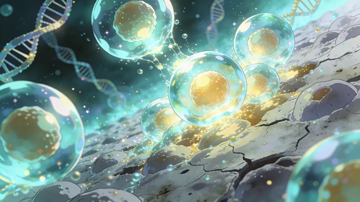
Autophagy: The cellular cleanup system behind healthy aging
Autophagy recycles damaged proteins and organelles. Learn what human research supports, how fasting and exercise may help, and what to track cautiously.
In 2016, Yoshinori Ohsumi won the Nobel Prize in Physiology or Medicine for a discovery most people have never heard of. He identified the genes and mechanisms behind autophagy — the process by which your cells break down and recycle their own damaged components. It was the scientific validation of something our bodies have been doing for millions of years: cleaning house at the molecular level to stay alive.
Since that Nobel Prize, autophagy research has exploded, and longevity scientists consistently rank it among the most important maintenance systems in aging biology. The evidence is strongest in cells and model organisms: when autophagy falters, damaged proteins accumulate, dysfunctional mitochondria persist, and molecular debris can amplify inflammation and several hallmarks of aging. Human research is moving quickly, but translating those mechanisms into reliable interventions is still a work in progress.
The useful takeaway is not that you can flip autophagy on at will. It is that exercise, metabolic health, sleep, nutrient timing, and diet quality can support the broader conditions that healthy cellular cleanup depends on.
What you’ll learn:
- What autophagy is and how it works at the cellular level
- Why autophagy declines with age and what that means for your health
- The three main types of autophagy and which one matters most for longevity
- 7 evidence-informed strategies that may support healthy autophagy signaling
- How autophagy interacts with mTOR and AMPK to determine your biological aging rate
Quick answer
Autophagy is the regulated recycling process cells use to break down damaged proteins, worn organelles, and cellular debris. It matters for longevity because weaker autophagy can worsen proteostasis, mitochondrial quality control, inflammation, and tissue repair. Fasting, exercise, sleep regularity, and nutrient quality may support autophagy, but human evidence is still more indirect than the strongest animal data.
Key facts
- Autophagy degrades damaged proteins and organelles.
- mTOR suppresses autophagy when nutrients and insulin are abundant.
- AMPK promotes autophagy during cellular energy stress.
- Human autophagy is hard to measure outside research settings.
- SuperAge tracks proxy signals, not a direct autophagy score.
What is autophagy?
The word autophagy comes from the Greek auto (self) and phagein (to eat). Literally: self-eating. But that dramatic name describes something elegantly constructive — your cells systematically identifying damaged, misfolded, or unnecessary components and breaking them down into raw materials that can be reused to build new, functional structures.
Quick definition: Autophagy is a highly regulated cellular process that degrades and recycles damaged organelles, misfolded proteins, and cellular debris to maintain cellular health and energy balance.
Think of autophagy like a city’s waste management and recycling system. Every day, your cells generate molecular waste — damaged mitochondria that leak reactive oxygen species, proteins that have lost their proper shape, and cellular membranes that no longer function correctly. Without a cleanup crew, this debris accumulates, interferes with normal operations, and eventually poisons the environment. Autophagy is that crew: it collects the waste, breaks it into useful raw materials, and feeds those materials back into the construction pipeline.
Why autophagy matters for your health
Autophagy isn’t just cellular housekeeping — it’s a fundamental survival mechanism with far-reaching consequences:
- Neurodegeneration prevention: Defective autophagy allows toxic protein aggregates to accumulate in neurons, contributing to Alzheimer’s, Parkinson’s, and Huntington’s disease
- Cancer suppression: Autophagy removes damaged DNA and dysfunctional organelles before they can trigger malignant transformation
- Immune function: Autophagy helps immune cells clear intracellular pathogens and present antigens for adaptive immune responses
- Metabolic regulation: The recycled amino acids and fatty acids from autophagy provide alternative fuel during energy stress
- Inflammation control: By removing damaged mitochondria (a process called mitophagy), autophagy prevents the release of inflammatory molecules that drive chronic low-grade inflammation
When autophagy works efficiently, your cells remain clean, functional, and resilient. When it fails, the consequences cascade through every organ system in your body. Autophagy decline is now formally recognized as one of the 12 hallmarks of aging — the primary biological mechanisms that drive the aging process.
The science behind autophagy
Understanding how autophagy actually works reveals why it’s so central to aging — and why activating it correctly matters more than simply “fasting for 16 hours.”
The five steps of autophagy
Autophagy follows a precise, multi-step sequence:
- Initiation: Stress signals — nutrient deprivation, energy depletion, or cellular damage — activate the ULK1 complex, which begins the autophagy cascade
- Nucleation: A structure called the phagophore begins to form, creating a double-membrane cup that will engulf the targeted cellular debris
- Expansion: The phagophore extends around the cargo — damaged mitochondria, protein aggregates, or invading pathogens — eventually sealing to form a complete autophagosome
- Fusion: The autophagosome merges with a lysosome — an organelle packed with digestive enzymes — creating an autolysosome
- Degradation and recycling: Lysosomal enzymes break down the cargo into amino acids, fatty acids, and nucleotides that are released back into the cytoplasm for reuse
This entire process takes 15–20 minutes per autophagosome, and a single cell can form hundreds simultaneously when the signal is strong enough.
The three types of autophagy
Not all autophagy is created equal. Your cells use three distinct mechanisms depending on what needs to be cleared:
| Type | Target | Mechanism | Relevance to Aging |
|---|---|---|---|
| Macroautophagy | Organelles, protein aggregates, pathogens | Double-membrane autophagosome engulfs cargo | Most studied; primary longevity target |
| Microautophagy | Small cytoplasmic components | Lysosome directly engulfs material through membrane invagination | Continuous baseline cleanup |
| Chaperone-mediated autophagy (CMA) | Specific damaged proteins with KFERQ motif | Chaperone proteins guide individual targets to the lysosome | Declines most dramatically with age |
When longevity researchers discuss autophagy, they’re primarily referring to macroautophagy — the large-scale cellular cleanup that responds to fasting, exercise, and metabolic stress. But chaperone-mediated autophagy deserves special attention: a landmark 2025 study in Nature Metabolism confirmed that CMA declines in aging human skeletal muscle, driving impaired calcium handling and mitochondrial dysfunction — mechanisms that directly contribute to age-related myopathy. The decline is primarily driven by reduced stability of LAMP2A (the rate-limiting CMA receptor) at the lysosomal membrane, caused by age-related changes in lysosomal lipid composition.
The master regulator: TFEB
Above the day-to-day autophagy machinery sits TFEB (Transcription Factor EB) — a master transcription factor that orchestrates both lysosomal biogenesis and autophagy gene expression. When cells need more cleanup capacity, TFEB moves into the nucleus and binds CLEAR motifs on dozens of autophagy and lysosomal genes simultaneously, effectively scaling up the entire system. A 2025 review in Frontiers in Bioscience identified TFEB (together with FOXO transcription factors) as one of the most promising therapeutic targets for age-related autophagy decline — its activity drops with age, and restoring it rescues multiple hallmarks of aging simultaneously, including proteostasis loss, mitochondrial dysfunction, and cellular senescence.
How autophagy affects your body
Autophagy operates in every tissue, but its effects are most dramatic in organs with high metabolic demands:
Brain: Neurons are post-mitotic — they don’t divide and replace themselves. This makes autophagy one of their key mechanisms for clearing accumulated protein waste. A 2010 study in Nature showed that mice with neuron-specific autophagy deficiency developed progressive neurodegeneration, including the accumulation of ubiquitinated protein inclusions similar to those found in Alzheimer’s disease. Human neurodegenerative disease is more complex, but impaired autophagy is now considered one important contributor to proteostasis failure and neuroinflammation.
Liver: Hepatocytes rely on autophagy to regulate lipid metabolism. Impaired hepatic autophagy contributes to metabolic dysfunction-associated steatotic liver disease (MASLD, formerly NAFLD), now affecting roughly 30% of the global population.
Heart: Cardiac muscle cells need constant autophagy to clear damaged mitochondria. When cardiac autophagy fails, it leads to cardiomyopathy and accelerated heart failure.
Skeletal muscle: Autophagy maintains muscle fiber quality by removing damaged contractile proteins. Its decline contributes directly to sarcopenia — the age-related loss of muscle mass and strength. The 2025 Nature Metabolism study linked falling CMA activity in aging muscle directly to progressive myopathy in both mice and humans.
Autophagy and longevity: what the research says
The connection between autophagy and lifespan is strong in model organisms and increasingly relevant to human aging biology, but human intervention evidence remains early:
- Genetic evidence: In model organisms such as C. elegans, altering autophagy genes can change lifespan, and blocking autophagy can blunt some benefits of dietary restriction
- Pharmacological evidence: Rapamycin extends lifespan in mice through mTOR inhibition, with autophagy likely contributing alongside other pathways. A 2025 PNAS paper introduced AA-20, a small-molecule autophagy activator that extended C. elegans lifespan and cleared protein aggregates without inhibiting mTORC1 — promising, but still preclinical
- Human evidence: human studies can measure some autophagy-related markers in blood or tissue, but there is no validated consumer test showing that a lifestyle routine has extended lifespan through autophagy
- Exercise data: animal studies show exercise benefits can depend partly on autophagy genes; in humans, exercise is best supported for fitness, insulin sensitivity, muscle, and inflammation, with autophagy as a plausible mechanism
The critical insight is cautious but important: autophagy is a major maintenance pathway, and many longevity interventions touch it, but at-home claims should not jump from mechanism to guaranteed human lifespan extension.
Why autophagy declines with age
If autophagy is so protective, why does it weaken precisely when you need it most?
Several age-related changes conspire to suppress autophagy:
1. Chronic mTOR activation
The mTOR signaling pathway is autophagy’s master suppressor. When mTOR is active — sensing abundant nutrients, amino acids, and insulin — it directly phosphorylates and inhibits ULK1, the kinase that initiates autophagy. Modern lifestyles, with constant food availability and high protein intake, keep mTOR chronically elevated, leaving autophagy perpetually suppressed.
2. AMPK decline
AMPK is autophagy’s primary activator. When cellular energy drops, AMPK activates ULK1 and inhibits mTOR, creating a powerful pro-autophagy signal. But AMPK responsiveness declines with age — the sensor becomes less sensitive to energy stress, meaning older cells need a stronger signal to trigger the same autophagic response.
3. Lysosomal dysfunction
Even when autophagosomes form correctly, they need functional lysosomes to complete degradation. Aging lysosomes accumulate an indigestible pigment called lipofuscin — the “age pigment” visible as brown spots on aging skin. Lipofuscin-loaded lysosomes lose their degradative capacity, creating a bottleneck where autophagosomes queue up but can’t be processed. Recent research also identifies altered lysosomal membrane lipid composition as a key driver of LAMP2A instability — the same mechanism that impairs chaperone-mediated autophagy with age.
4. Transcriptional changes
The expression of key autophagy genes — including ATG5, ATG7, and BECN1 — decreases with age in multiple tissues, driven in part by reduced TFEB and FOXO transcription factor activity. Animal studies show that restoring expression of just one of these genes (ATG5) in aging mice improves motor function, metabolism, and lifespan.
5. NAD+ depletion
NAD+ (nicotinamide adenine dinucleotide) is required for sirtuin activation, and sirtuins (particularly SIRT1) interact with autophagy-related pathways. NAD+ biology changes with age and metabolic stress, but supplement-driven autophagy claims in humans remain more uncertain than the cellular mechanisms.
The result is a vicious cycle: declining autophagy leads to accumulated cellular damage, which further impairs autophagic machinery, accelerating the decline. Breaking this cycle is one of the most promising strategies in longevity science.
7 evidence-informed strategies that may support autophagy
Evidence note
Most lifestyle claims about autophagy come from animal studies, cell biology, or indirect human markers. Use fasting windows, protein timing, sauna, cold exposure, supplements, and wearable data as practical levers to discuss with a clinician when relevant, not as proof that autophagy is “on” or that biological aging has reversed.
1. Time-restricted eating and fasting windows
Why it works: Fasting windows can lower insulin exposure, reduce nutrient signaling through mTOR, and increase energy-stress signals such as AMPK. A 2025 randomized trial in The Journal of Physiology measured autophagic flux markers in peripheral blood cells from adults with obesity after 6 months of intermittent time-restricted eating and found a modest increase compared with standard care. That is useful human evidence, but it does not prove that every 16-hour fast produces whole-body autophagy or longevity benefits.
How to do it:
- Start with a 16:8 intermittent fasting protocol (16 hours fasting, 8 hours eating)
- Narrow the eating window only if sleep, training, mood, and glucose control remain stable
- Be cautious with longer fasts if you are lean, older, pregnant, highly active, have an eating-disorder history, or use glucose-lowering medication
- Black coffee and plain tea are compatible with many fasting protocols, but they are not proven human autophagy enhancers
What to expect: Some people see better weight, insulin, triglycerides, and evening glucose patterns over weeks to months. Direct autophagy measurement is still research-grade and tissue-specific.
2. High-intensity exercise
Why it works: Exercise raises cellular energy demand, activates AMPK-related signaling, and stimulates repair pathways in muscle and other tissues. A landmark 2012 Nature mouse study showed that exercise-induced autophagy was needed for some metabolic benefits of physical activity. In humans, exercise is best supported through outcomes such as fitness, insulin sensitivity, muscle function, and inflammation.
How to do it:
- Build aerobic exercise into most weeks, from brisk walking to cycling, swimming, or running
- Use HIIT protocols if your joints, heart risk, and recovery make them appropriate
- Resistance training also activates autophagy through mechanical stress — aim for 2–3 sessions per week
- Fasted exercise is optional; performance, safety, and consistency matter more than stacking stressors
What to expect: Fitness, insulin sensitivity, resting heart rate, HRV, and strength can improve over weeks to months. Those changes support the broader cellular-repair environment without proving a specific autophagy dose.
3. Protein timing without underfeeding
Why it works: Amino acids — particularly leucine — activate mTOR, which can suppress autophagy while supporting muscle protein synthesis. The practical goal is not chronic low protein; it is cycling feeding and fasting signals while preserving muscle, especially after 40.
How to do it:
- On 5–6 days per week, consume adequate protein for muscle maintenance: 0.7–1.0 g/lb (1.6–2.2 g/kg) body weight
- Avoid constant snacking if it drives excess calories or poor glucose control
- Put most protein in meals rather than grazing all day
- Do not use low-protein days if you are frail, underweight, recovering from illness, pregnant, or trying to build muscle aggressively
What to expect: Protein timing may help balance mTOR-linked growth and fasting-linked repair signals, but direct human evidence for scheduled low-protein days increasing beneficial autophagy is limited.
4. Spermidine-rich foods
Why it works: Spermidine is a natural polyamine that induces autophagy in many experimental models. A 2024 Nature Cell Biology paper showed that spermidine was important for fasting-mediated autophagy and longevity in model systems. Human data are still mostly observational: higher dietary spermidine intake has been associated with lower mortality, but that does not prove supplementation extends human lifespan. The ongoing POLYCAD trial is testing 24 mg/day spermidine for 48 weeks in 187 adults aged 65+ with coronary artery disease, with outcomes including cardiac remodeling, VO2 peak, lean mass, and hs-CRP.
How to do it:
- Prioritize spermidine-rich foods: wheat germ (the richest source at 24 mg/100g), aged cheese (particularly cheddar and parmesan), mushrooms, soybeans, legumes, and green peas
- Include 1–2 tablespoons of wheat germ daily in smoothies, yogurt, or oatmeal
- Fermented foods (natto, miso) provide both spermidine and other pro-autophagic compounds
- Treat supplements cautiously until stronger clinical outcome data are available, especially if you have cancer history, kidney disease, pregnancy, or complex medications
What to expect: Spermidine-rich foods can be part of a nutrient-dense pattern. Whether they meaningfully change autophagy markers or cardiovascular outcomes in an individual human is not yet predictable.
5. Heat and cold stress
Why it works: Thermal stress activates heat-shock and cold-shock responses that overlap with proteostasis and autophagy biology. Sauna bathing has stronger human evidence for cardiovascular and heat-stress adaptation than for direct autophagy measurement. Cold exposure can activate brown adipose tissue and AMPK-related pathways, but autophagy-specific evidence in humans remains limited.
How to do it:
- Sauna sessions: 15–20 minutes at 176–212°F (80–100°C), 3–4 times per week
- Cold exposure: 2–5 minutes of cold water immersion at 50–59°F (10–15°C), or end showers with 60–90 seconds of cold water
- Hydrate well and avoid heat or cold exposure when ill, dehydrated, intoxicated, or medically unstable
- Treat contrast therapy as optional; additive autophagy benefits are not established in humans
What to expect: Regular sauna may support cardiovascular markers and perceived recovery. Cold exposure may improve cold tolerance and alertness. Neither should replace exercise, sleep, nutrition, or medical care.
6. Sleep optimization
Why it works: Autophagy and lysosomal function follow circadian rhythms, and sleep supports brain waste-clearance systems, immune regulation, and metabolic recovery. Poor sleep is linked with worse glucose control, higher appetite, and impaired recovery; direct claims about a specific nightly autophagy dose in humans are still premature.
How to do it:
- Prioritize 7–9 hours of total sleep and a consistent sleep-wake schedule
- Keep the bedroom dark and cool — 65–68°F (18–20°C) is optimal
- Avoid heavy late meals if they worsen sleep or glucose patterns
What to expect: Better sleep can improve recovery, appetite regulation, glucose control, and cognitive clarity. These are useful proxy outcomes, not proof that brain autophagy has increased.
7. Polyphenol-rich diet
Why it works: Several plant polyphenols influence autophagy-related pathways in cell and animal models. Resveratrol, EGCG, curcumin, oleocanthal, and sulforaphane interact with nutrient-sensing, oxidative-stress, and inflammatory pathways, but human effects depend on dose, food matrix, gut microbiome, baseline health, and adherence.
How to do it:
- Drink 2–3 cups of green tea daily (rich in EGCG)
- Include deeply colored berries, particularly blueberries and blackberries, with most meals — pomegranates, raspberries, and walnuts also provide ellagitannins that gut bacteria convert to urolithin A, a potent mitophagy activator with clinical trial evidence
- Use turmeric with black pepper in cooking if you tolerate it
- Consume extra virgin olive oil daily — oleocanthal activates autophagy pathways
- Dark chocolate (85%+ cacao) provides catechins and epicatechins that support autophagic flux
What to expect: A polyphenol-rich diet is most useful as part of an overall dietary pattern that improves inflammation, vascular health, and metabolic markers. It is not a stand-alone autophagy protocol.
How to track and measure autophagy
Unlike cholesterol or blood glucose, you can’t directly measure autophagy with a standard blood test. Several indirect biomarkers and functional indicators can tell you whether your metabolic environment is more compatible with repair and recovery, but they do not prove tissue-level autophagy is active.
Key metrics to monitor
| Metric | How to Measure | What It Indicates |
|---|---|---|
| Fasting insulin | Blood test (fasted) | Lower-normal values, interpreted with glucose and body composition, suggest better insulin sensitivity |
| Glucose and ketone trends | CGM, fingerstick glucose, or ketone meter | Can show whether fasting or lower-carb periods shift fuel use, but do not directly quantify autophagy |
| hs-CRP | Blood test | Lower values outside infection or injury suggest lower systemic inflammation |
| Blood ketones (BHB) | Ketone meter | Indicates a metabolic shift toward fat oxidation; autophagy effects remain inferred |
| Deep sleep duration | Sleep tracker / Apple Watch | Helps track recovery and circadian consistency, not a direct nocturnal autophagy window |
| HRV (heart rate variability) | Wearable device | Higher or improving HRV can reflect better autonomic recovery and stress tolerance |
Indirect signs of active autophagy
When your broader recovery pattern is improving, you may notice:
- Mental clarity: fewer brain-fog episodes, especially when sleep and glucose patterns improve
- Stable energy: fewer mid-afternoon crashes and more consistent training readiness
- Better exercise recovery: quicker bounce-back from workouts, with less persistent soreness
- Lower inflammatory markers: declining hs-CRP or improved NLR over time, if other causes are excluded
These signs are useful, but they are not specific autophagy measurements.
How SuperAge helps you optimize autophagy
Important: SuperAge does not measure autophagy directly. It organizes sleep, HRV, activity, metabolic, and lab trends that may sit upstream or downstream of autophagy, so the app is best used for pattern recognition rather than as an autophagy diagnostic test.
Tracking autophagy’s indirect markers manually — fasting insulin, deep sleep, HRV, inflammatory markers — requires juggling multiple apps and lab results. SuperAge brings these signals together in one place.
Metabolic health monitoring
SuperAge tracks key metabolic and recovery indicators through Apple Health integration, including resting heart rate, heart rate variability, activity patterns, sleep, and lab trends. These signals sit near the lifestyle inputs that influence autophagy biology, but they do not diagnose autophagy activation. For the complete picture of how metabolic health and cellular maintenance interact with aging, see our metabolic health and aging guide.
Sleep quality analysis
By monitoring your sleep stages through Apple Watch data, SuperAge helps you understand whether your sleep timing and recovery are improving. You can see trends over weeks and months, identifying patterns that either support or undermine the recovery environment cellular cleanup depends on.
Your biological age, tracked
Autophagy is one maintenance pathway among many that influence aging biology. SuperAge’s biological age calculation captures downstream health signals such as inflammation, metabolic health, cardiovascular fitness, and body composition. As you implement fasting, exercise, sleep, and dietary strategies, track whether those measurable inputs improve rather than assuming an autophagy score has changed.
Frequently asked questions
How long do you need to fast to trigger autophagy?
Human research is still evolving, and there is no universal hour-count that proves autophagy has turned on across your tissues. Animal studies suggest fasting can raise autophagy markers, while a 2025 human trial found that six months of intermittent time-restricted eating increased autophagic flux markers in blood cells compared with standard care. A consistent 12- to 16-hour overnight fast may be a practical starting point for metabolic health, but longer fasts should be individualized and medically supervised when risk factors exist.
Can you activate autophagy without fasting?
Yes, fasting is not the only lever. Exercise, sleep regularity, nutrient-dense diets, and metabolic health all influence pathways that overlap with autophagy biology. Polyphenol-rich diets, heat stress, and cold exposure are plausible supporting tools, but human evidence is more indirect than the mechanism diagrams make it look.
Does autophagy fight cancer or promote it?
Autophagy has a dual role in cancer. In healthy tissue it can help remove damaged components that may contribute to tumor initiation, while established tumors can sometimes use autophagy to survive stress or resist treatment. If you have active cancer, a cancer history, or are in treatment, discuss fasting, supplements, or deliberate autophagy-targeting with your oncology team first.
Does coffee break autophagy during a fast?
Plain black coffee is unlikely to stop a fast in the same way a caloric drink would, and coffee compounds have induced autophagy in animal models. Direct human data is limited, so it is safer to say that black coffee is compatible with many fasting protocols rather than a proven autophagy enhancer. Milk, cream, sugar, and calorie-containing additives change the metabolic signal.
At what age does autophagy start declining?
Autophagy appears to become less efficient with age, but there is no single birthday when it “starts declining” for everyone. Different tissues and autophagy pathways change at different rates, and human measurement remains indirect. Consistent exercise, adequate sleep, metabolic health, and nutrient quality are more actionable than trying to pinpoint an exact age threshold.
Key takeaways
- Autophagy is cellular recycling: it breaks down damaged proteins, organelles, and debris so cells can reuse the raw materials.
- Aging and autophagy interact: weaker cleanup can contribute to proteostasis loss, mitochondrial dysfunction, inflammation, and tissue fragility.
- mTOR and AMPK shape the signal: abundant nutrients tend to suppress autophagy, while energy stress tends to promote it.
- Human evidence needs caution: fasting, exercise, spermidine-rich foods, thermal stress, sleep, and polyphenols are promising but not direct at-home autophagy switches.
- Tracking should be indirect: combine sleep, HRV, activity, metabolic labs, and symptoms to see whether the broader recovery pattern is improving.
Start your cellular renewal journey today
Autophagy is happening in your body right now, but it is not something you can score with a wearable or switch on with one habit. The practical target is a recovery-friendly routine: train consistently, sleep well, eat nutrient-dense food, avoid constant overfeeding, and use fasting windows only when they fit your physiology and life.
Ready to take control? Download SuperAge and start tracking the metabolic, sleep, activity, and recovery markers that reflect your broader cellular-maintenance environment — alongside your biological age.
References
- The Nobel Prize in Physiology or Medicine 2016: Yoshinori Ohsumi
- Autophagy as a promoter of longevity: insights from model organisms
- Autophagy and the cell biology of age-related disease
- Intermittent time-restricted eating may increase autophagic flux in humans
- Age-related decline of chaperone-mediated autophagy in skeletal muscle leads to progressive myopathy
- Spermidine is essential for fasting-mediated autophagy and longevity
- POLYCAD trial: spermidine in older adults with coronary artery disease
- Autophagy activator AA-20 and proteostasis in C. elegans
- Transcriptional dysregulation of autophagy in aging: TFEB and FOXOs
- Autophagy in cancer: context-dependent tumor suppression and tumor support
- Links between autophagy and healthy aging. Journal of Molecular Biology. 2026.
Last updated: 2026-07-07. This article is regularly reviewed to ensure accuracy.
Related Articles
mTOR: When to turn it off to live longer
mTOR drives cell growth but accelerates aging when chronically active. Learn how mTOR works, why inhibition extends lifespan in animal studies, and 7 natural ways to optimize it.