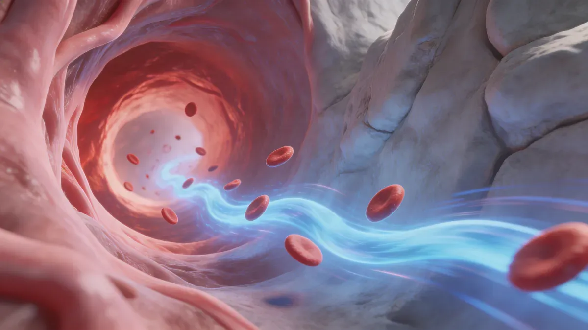
Aortic stiffness and pulse wave velocity: The gold standard of vascular age
Learn how pulse wave velocity reflects aortic stiffness and vascular age, what values mean, how testing works, and which habits may improve arterial health.
Quick answer
Pulse wave velocity (PWV) estimates how fast the pressure wave from each heartbeat travels through the aorta and large arteries. Faster values usually mean stiffer arteries and higher cardiovascular risk. Carotid-femoral PWV is the reference non-invasive measure of aortic stiffness, but results should be interpreted with age, blood pressure, device type, measurement method, and overall cardiovascular risk.
Key facts
- Pulse wave velocity | measures | arterial pressure-wave speed, not plaque burden or cholesterol directly.
- Carotid-femoral PWV | is | the reference non-invasive test for aortic stiffness in clinical research and many cardiology settings.
- Higher PWV | is associated with | higher cardiovascular event and mortality risk, including in meta-analyses that adjust for traditional risk factors.
- Vascular age tracking | works best | when PWV is interpreted alongside blood pressure, pulse pressure, ApoB, glucose, inflammation, fitness, and kidney health.
Your blood pressure might be 120/80. Your cholesterol might be perfect. You might even exercise five days a week. But there’s a number your doctor may never have measured - one that can add risk information beyond traditional factors: the speed at which pressure waves travel through your aorta.
It’s called pulse wave velocity (PWV), and carotid-femoral PWV is the reference non-invasive measurement of aortic stiffness - one important structural change in cardiovascular aging. When your aorta is young and elastic, pressure waves travel slowly: around 6 m/s (20 ft/s). As it stiffens with age, those waves accelerate - sometimes reaching 10-12 m/s (33-39 ft/s) or more by your 60s and 70s.
In one meta-analysis, every 1 m/s (3.3 ft/s) higher aortic PWV was associated with about 14% higher risk of cardiovascular events and 15% higher all-cause mortality after adjustment for age, sex, and traditional risk factors. Yet this measurement takes about 10-15 minutes, requires no needles or radiation, and is available in many cardiology clinics today.
What you’ll learn:
- What pulse wave velocity measures and why it’s the gold standard
- The physics and biology of aortic stiffening
- Normal PWV values by age and how to interpret your results
- How PWV testing works
- 7 evidence-based strategies to reduce arterial stiffness
- How to track vascular aging with biomarkers
What is pulse wave velocity?
Every time your heart beats, it creates a pressure wave that travels from the aorta through your arterial tree to the peripheral vessels. Pulse wave velocity is the speed of that wave — measured in meters per second (m/s).
Quick definition: Pulse wave velocity (PWV) is the speed at which the arterial pressure wave generated by each heartbeat travels through the aorta and large arteries. Carotid-femoral PWV is the reference non-invasive measurement of aortic stiffness and an independent predictor of cardiovascular events and mortality.
Why stiffness determines speed
The physics is simple: pressure waves travel faster through rigid materials and slower through elastic ones. Think of tapping one end of a steel pipe versus a rubber hose — the vibration reaches the other end much faster through steel.
Your aorta works the same way:
- Young, elastic aorta (lots of elastin, compliant wall): PWV ~6 m/s (20 ft/s) → pressure wave arrives slowly → organs receive smooth, buffered blood flow
- Aged, stiff aorta (fragmented elastin, excess collagen, calcification): PWV ~12 m/s (39 ft/s) → pressure wave arrives rapidly → organs receive pulsatile, damaging blood flow
Why this matters for aging
A stiff aorta doesn’t just passively reflect aging — it actively drives it. The Windkessel function of the aorta (its ability to absorb the heart’s ejection pressure and release it smoothly during diastole) is essential for protecting two critical organ systems:
-
The heart: A stiff aorta reflects pressure waves back to the heart during systole (instead of diastole), increasing cardiac workload and causing left ventricular hypertrophy — the thickening of the heart muscle that precedes heart failure.
-
The microcirculation: High-flow organs — especially the brain and kidneys — are exquisitely sensitive to pulsatile flow. When the aorta fails to buffer pressure, these organs receive damaging pulses that accelerate cognitive decline, kidney disease, and microvascular damage.
This is why aortic stiffness is increasingly studied as both a marker and a contributor to cardiovascular aging.
The science behind aortic stiffening
The structural transformation
The aortic wall undergoes specific structural changes with age that directly increase PWV:
Elastin fragmentation: Elastin is the protein that gives the aorta its rubber-band-like properties. Unlike collagen, elastin has virtually zero turnover in adulthood — the elastin in your aorta today is the same elastin that was synthesized during fetal development. Over decades, mechanical stress (3 billion heartbeats by age 80) progressively fragments these irreplaceable fibers.
Collagen deposition: As elastin degrades, the body compensates by producing collagen — a much stiffer structural protein. The collagen-to-elastin ratio increases progressively, shifting the aortic wall from elastic to rigid.
Glycation cross-links: Advanced glycation end products (AGEs) form cross-links between collagen fibers, making them even more rigid. This process accelerates with elevated blood sugar — explaining why diabetes is one of the strongest accelerators of arterial stiffening.
Medial calcification: Calcium deposits in the medial layer of the aorta (Mönckeberg sclerosis) create rigid segments that dramatically increase local PWV. Unlike atherosclerotic calcification (which occurs in the intima), medial calcification is primarily age-related and strongly correlates with coronary calcium scores.
Endothelial dysfunction: When the endothelium stops producing adequate nitric oxide, smooth muscle cells in the arterial wall remain chronically contracted — a functional component of stiffness that is potentially reversible.
The acceleration after 50
PWV increases approximately 6-8% per decade throughout life, but this rate accelerates markedly after age 50. The inflection point coincides with:
- Critical depletion of elastin reserves
- Accumulated glycation damage
- Onset of medial calcification
- Hormonal changes (menopause in women dramatically accelerates aortic stiffening)
- Cumulative exposure to blood pressure, inflammation, and metabolic stress
PWV and the hallmarks of aging
Aortic stiffness connects to multiple hallmarks of aging:
- Loss of proteostasis: Elastin cannot be replaced; damaged collagen accumulates
- Cellular senescence: Senescent vascular smooth muscle cells secrete inflammatory mediators that promote stiffening
- Altered intercellular communication: The failing endothelium-smooth muscle signaling axis drives chronic vasoconstriction
- Epigenetic alterations: Epigenetic changes in vascular cells alter gene expression patterns that maintain arterial compliance
Normal PWV values by age
Reference ranges (carotid-femoral PWV)
| Age Group | Normal Range (m/s) | Normal Range (ft/s) | Median | 90th Percentile |
|---|---|---|---|---|
| 20-29 | 5.5-7.0 | 18-23 | 6.2 | 7.6 |
| 30-39 | 5.8-7.5 | 19-25 | 6.5 | 8.0 |
| 40-49 | 6.5-8.5 | 21-28 | 7.2 | 9.8 |
| 50-59 | 7.0-10.0 | 23-33 | 8.3 | 12.1 |
| 60-69 | 8.0-12.0 | 26-39 | 10.3 | 15.0 |
| 70-79 | 9.0-14.0 | 30-46 | 11.5 | 16.0+ |
Clinical thresholds
European hypertension guidance and arterial-stiffness consensus documents have used these practical cutoffs, but age, blood pressure at the time of testing, device type, and measurement method all matter:
- < 10 m/s (33 ft/s): Often considered lower-risk in many adults, especially when blood pressure is controlled
- 10-12 m/s (33-39 ft/s): Elevated for many middle-aged adults; interpret against age-specific reference ranges
- > 12 m/s (39 ft/s): High in many clinical contexts; worth discussing with a clinician if persistent
- > 14 m/s (46 ft/s): Very high; often seen with uncontrolled hypertension, diabetes, chronic kidney disease, or advanced vascular aging
Calculating your “vascular age”
One way to estimate vascular age is to compare your PWV with the age-specific median from reference populations:
- A 45-year-old with PWV of 9.5 m/s (31 ft/s) has a vascular age of approximately 58
- A 65-year-old with PWV of 8.0 m/s (26 ft/s) has a vascular age of approximately 50
This gap between chronological and vascular age can be useful in longevity medicine, especially when it is tracked alongside blood pressure, ApoB, glucose, kidney markers, inflammation, and fitness.
How PWV testing works
Carotid-femoral PWV (the gold standard)
The reference standard measurement assesses aortic stiffness by measuring the speed of the pressure wave between the carotid artery (neck) and the femoral artery (groin):
- Two pressure sensors (tonometers or cuffs) are placed: one on the neck over the carotid artery, one on the groin over the femoral artery
- The device records the pressure waveform at both sites simultaneously
- PWV is calculated: distance between sensors ÷ transit time of the pressure wave
- The result is expressed in meters per second (m/s)
- Total time: 10-15 minutes
Other measurement methods
| Method | What It Measures | Accessibility | Accuracy |
|---|---|---|---|
| Carotid-femoral PWV | Aortic stiffness (gold standard) | Cardiology clinics | Highest |
| Brachial-ankle PWV (baPWV) | Combined aortic + peripheral stiffness | Many clinics (common in Asia) | Good (slightly overestimates) |
| Oscillometric devices | Estimated PWV from blood pressure cuff | Office-based, automated | Moderate |
| CIMT + PWV | Structural + functional vascular assessment | Specialized centers | Complementary |
| MRI-based PWV | Direct aortic measurement with imaging | Research/hospitals | Excellent but expensive |
Preparation for testing
- Avoid caffeine for 3 hours before testing
- No smoking for 3 hours before
- Rest in supine position for 10 minutes before measurement
- Empty bladder (full bladder can increase sympathetic tone)
- Avoid heavy meals 2 hours before
- Measurements should be taken in a quiet, temperature-controlled room
7 evidence-informed strategies to reduce arterial stiffness
1. Aerobic exercise: one of the best-supported interventions
Why it works: Chronic aerobic exercise is one of the best-supported interventions for improving PWV. It improves endothelial function, increases nitric oxide production, reduces chronic inflammation, and may partially restore functional arterial compliance even when structural changes are present.
How to do it:
- Aim for 150-300 minutes of moderate-intensity aerobic exercise per week
- Zone 2 training (60-70% max HR) is particularly effective for vascular benefits
- Walking at brisk pace (3.5+ mph / 5.6+ km/h) significantly reduces PWV
- Swimming provides excellent vascular benefits due to hydrostatic pressure effects
- Do not rely only on very heavy resistance training if PWV is a concern; discuss programming with a clinician or qualified coach if you have hypertension or cardiac risk
Expected results: Studies of exercise training often report clinically meaningful PWV reductions, though the exact change depends on baseline stiffness, blood pressure, age, training type, and measurement method.
2. Reduce sodium and increase potassium
Why it works: Sodium directly stiffens arteries independent of blood pressure — even in people whose blood pressure doesn’t respond to salt. Sodium enters endothelial cells and stiffens their glycocalyx (surface layer), impairing nitric oxide release and increasing vascular tone. Potassium has the opposite effect, relaxing arterial smooth muscle and promoting sodium excretion.
How to do it:
- Target < 2,300 mg sodium/day, or a lower target if your clinician recommends it
- Increase potassium-rich foods such as sweet potatoes, bananas, spinach, avocados, and beans; use caution with potassium targets if you have kidney disease or take ACE inhibitors, ARBs, or potassium-sparing diuretics
- Emphasize whole foods over processed (80% of dietary sodium comes from processed foods)
- Use herbs and spices instead of salt for flavoring
Expected results: Some trials report PWV improvement with sodium reduction, but the effect varies and often overlaps with blood pressure change.
3. Optimize blood pressure
Why it works: Blood pressure and arterial stiffness form a bidirectional cycle: high blood pressure accelerates stiffening, and stiffened arteries increase blood pressure. Breaking this cycle is essential.
How to do it:
- Discuss an individualized blood pressure target; many appropriate adults benefit from lower, well-tolerated blood pressure, but targets depend on age, medications, frailty, kidney disease, and symptoms
- Blood pressure variability matters as much as average readings — high visit-to-visit variability independently increases PWV
- Monitor at home: morning measurements before medication are most revealing
- Address pulse pressure (systolic minus diastolic) — widening pulse pressure is the hallmark hemodynamic signature of aortic stiffness
Expected results: Blood pressure reduction often lowers PWV because it reduces pressure load on the arterial wall; longer-term benefit depends on sustained control.
4. Control glycation and blood sugar
Why it works: Glycation creates irreversible cross-links between collagen fibers in the arterial wall. These AGE cross-links are one of the few truly irreversible components of arterial stiffening (unlike functional stiffness from vasoconstriction, which is reversible). Preventing new cross-link formation is critical.
How to do it:
- Keep HbA1c in a healthy range for your context
- Monitor fasting glucose and post-meal patterns if you have insulin resistance or diabetes risk
- Minimize high-glycemic foods and added sugars
- Walk after meals to blunt glucose spikes
- Improve insulin sensitivity through exercise and weight management
Expected results: Better glucose control may slow cross-link formation and help stabilize PWV over time, especially when paired with exercise and weight management.
5. Adopt an anti-inflammatory dietary pattern
Why it works: Chronic inflammation promotes vascular smooth muscle proliferation, collagen deposition, and endothelial dysfunction — all of which increase arterial stiffness. The Mediterranean diet has the strongest evidence base for improving arterial compliance.
How to do it:
- Follow a Mediterranean dietary pattern: olive oil, fish, vegetables, legumes, nuts
- Eat omega-3 rich foods 3-4 times weekly (EPA/DHA improve arterial compliance directly)
- Include polyphenol-rich foods daily: berries, dark chocolate, green tea
- Increase fiber intake to 30+ g/day
- Minimize processed foods, refined sugars, and industrial seed oils
Expected results: Reduced hs-CRP, improved blood pressure, and better metabolic health may translate into better arterial compliance over time.
6. Manage weight and visceral fat
Why it works: Visceral fat secretes inflammatory adipokines and drives insulin resistance — both of which accelerate arterial stiffening. Even modest weight loss (5-10%) significantly reduces PWV. The effect is most pronounced when visceral fat is specifically reduced.
How to do it:
- Focus on body fat percentage rather than scale weight
- Combine resistance training with aerobic exercise (the combination is more effective for visceral fat reduction than either alone)
- Monitor waist circumference: < 35 in (89 cm) for women, < 40 in (102 cm) for men
- Address metabolic syndrome components comprehensively
Expected results: PWV may improve with 5-10% weight loss over 3-6 months, especially when visceral fat, blood pressure, and insulin resistance improve together.
7. Consider targeted nutritional support
Why it works: Several nutrients have limited but plausible evidence for supporting arterial compliance through distinct mechanisms - from enhancing nitric oxide production to supporting mineral balance.
How to do it:
- Magnesium: discuss dose and form if intake is low or you use blood pressure medication
- Vitamin K2: may support calcium handling, but avoid unsupervised use with warfarin
- Vitamin D: correct deficiency rather than chasing very high levels
- Beetroot juice or nitrate-rich foods may enhance nitric oxide and functional arterial compliance
- CoQ10: 100-200 mg/day (supports mitochondrial function in vascular smooth muscle cells)
The information provided does not replace professional medical advice. Consult your healthcare provider before starting any supplementation.
Expected results: Any PWV effect is usually modest and should be treated as secondary to exercise, blood pressure control, diet quality, sleep, and metabolic health.
Tracking vascular aging over time
The vascular aging battery
For comprehensive vascular age assessment, combine:
| Test | What It Measures | Recommended Frequency |
|---|---|---|
| Carotid-femoral PWV | Aortic stiffness (functional) | Every 1-2 years |
| CIMT | Arterial wall thickness (structural) | Every 1-2 years |
| Coronary calcium score | Calcified plaque | Every 3-5 years |
| Blood pressure | Hemodynamic stress | Continuous (wearable) |
| hs-CRP | Vascular inflammation | Every 6-12 months |
| ApoB | Atherogenic burden | Every 6-12 months |
Daily wearable metrics that reflect vascular health
Your Apple Watch can’t measure PWV directly, but it tracks metrics closely correlated with arterial stiffness:
- HRV: Higher HRV reflects better autonomic balance and lower vascular stiffness
- Resting heart rate: Lower RHR indicates efficient cardiovascular function
- Blood pressure trends: Rising systolic with stable diastolic = widening pulse pressure = sign of increasing aortic stiffness
- VO2 max: Strongly inversely correlated with PWV
How SuperAge monitors your vascular trajectory
Arterial stiffness is one important determinant of biological age - and SuperAge tracks the daily metrics that either accelerate or slow its progression.
Integrated cardiovascular monitoring
SuperAge combines HRV, resting heart rate, blood pressure trends, and VO2 max from your Apple Watch into a comprehensive cardiovascular profile. These metrics collectively reflect the vascular health that PWV measures directly.
Biological age as a vascular proxy
The PhenoAge algorithm used by SuperAge includes biomarkers (albumin, CRP, glucose) that correlate with arterial stiffness. Improvements in your biological age often parallel improvements in vascular function — giving you daily feedback between annual PWV measurements. For a full comparison of how biological age and vascular age relate — where they overlap and where they diverge — see our biological age vs vascular age guide.
Long-term trend analysis
SuperAge tracks your cardiovascular metrics over months and years, revealing whether your interventions are bending the vascular aging curve. This longitudinal view is invaluable for staying motivated between clinical vascular assessments.
Frequently asked questions
What’s the difference between PWV and arterial stiffness?
Arterial stiffness is the property; PWV is the measurement. Arterial stiffness refers to the reduced elasticity of artery walls. PWV quantifies this stiffness by measuring how fast pressure waves travel - faster waves mean stiffer arteries. Carotid-femoral PWV is the reference non-invasive measure because it directly reflects aortic compliance and has strong predictive value for cardiovascular events.
Can arterial stiffness be reversed?
Partially. The functional component (endothelial dysfunction, smooth muscle contraction, inflammation) is reversible with exercise, diet, and medication. The structural component (elastin fragmentation, glycation cross-links, calcification) is largely irreversible — but you can prevent further progression. In practice, most studies show PWV reductions of 0.5-1.5 m/s (1.6-5 ft/s) with lifestyle interventions — meaningful clinical improvements.
At what age should I get PWV tested?
Consider baseline testing at age 40-45 if you have any cardiovascular risk factors (hypertension, diabetes, family history, smoking). For those without risk factors, age 50 is a reasonable starting point. Earlier testing is appropriate if you’re pursuing proactive longevity medicine or have premature vascular aging indicators (widening pulse pressure, elevated CIMT).
How does PWV relate to blood pressure medications?
Antihypertensive treatment can reduce PWV, and much of the improvement may come from lowering mean arterial pressure itself. Some drug classes, including ACE inhibitors, ARBs, and calcium channel blockers, have been studied for arterial-stiffness effects, but medication choice depends on the whole clinical picture. If you’re on medication and interested in vascular aging, discuss these distinctions with your cardiologist.
Is high PWV the same as atherosclerosis?
No, though they’re related. Arterial stiffness (high PWV) is primarily a medial layer process — changes in collagen, elastin, and smooth muscle. Atherosclerosis is primarily an intimal layer process — lipid accumulation, plaque formation, and inflammation. Both contribute to cardiovascular risk, and they often coexist, but they can occur independently. CIMT better captures atherosclerosis, while PWV better captures stiffness.
Key takeaways
- cfPWV is the reference measure: Carotid-femoral pulse wave velocity is the most validated non-invasive measure of aortic stiffness and vascular aging
- Each 1 m/s increase carries signal: Meta-analyses link higher aortic PWV with higher cardiovascular-event and mortality risk, even after traditional risk adjustment
- The aorta contributes to aging: A stiff aorta can damage the heart, brain, and kidneys through pulsatile overload
- Intervention can help: Aerobic exercise, blood pressure control, diet quality, and metabolic health may improve or stabilize PWV
- Track context, not one number: Baseline PWV at 40-50, combined with daily wearable and biomarker trends, gives you a more actionable view of your vascular trajectory
Measure your vascular age — and change it
Your aorta is the main highway of your circulatory system. How fast pressure waves race through it determines how quickly your heart, brain, and kidneys age. The test takes 10 minutes. The interventions start today.
Ready to track the daily metrics that predict your vascular future? Download SuperAge and start monitoring HRV, resting heart rate, VO2 max, and your biological age — the numbers that tell you whether your arteries are aging faster or slower than expected.
References
- Laurent S et al. (2006). “Expert consensus document on arterial stiffness.” European Heart Journal, 27(21). — Gold standard PWV methodology and clinical thresholds.
- Ben-Shlomo Y et al. (2014). “Aortic pulse wave velocity improves cardiovascular event prediction.” Journal of the American College of Cardiology. — PWV predictive value meta-analysis.
- Vlachopoulos C et al. (2010). “Prediction of cardiovascular events and all-cause mortality with arterial stiffness.” European Heart Journal. — 14-15% risk per 1 m/s increase.
- Reference Values for Arterial Stiffness Collaboration (2010). “Determinants of pulse wave velocity in healthy people and in the presence of cardiovascular risk factors.” European Heart Journal. — Age-specific reference values.
- Ashor AW et al. (2014). “Effects of exercise modalities on arterial stiffness and wave reflection.” PLOS ONE. — Exercise intervention meta-analysis.
Last updated: 2026-03-19. This article is regularly reviewed to ensure accuracy.
Related Articles
Endothelial function: The vascular aging test your doctor doesn't order
Endothelial function shapes nitric oxide, blood flow, clotting, and vascular aging. Learn flow-mediated dilation testing and vessel-protective habits.