Proteostasis: How cellular quality control changes with age
Discover how loss of proteostasis contributes to aging — from misfolded proteins to neurodegenerative disease — and evidence-based ways to support protein quality control.
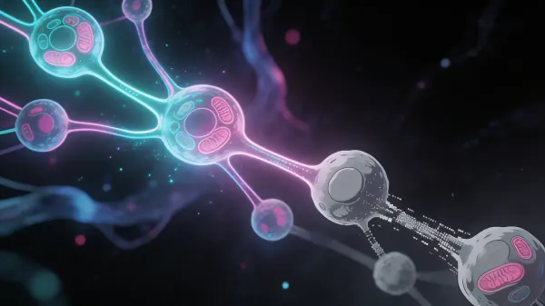
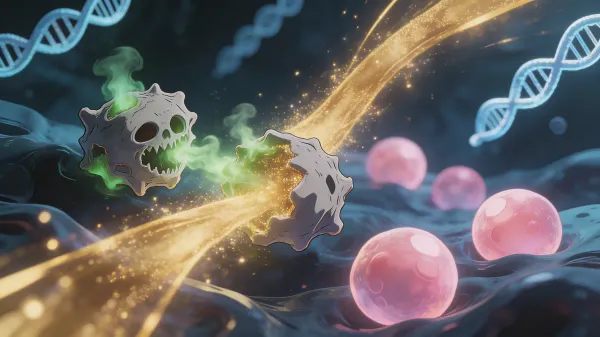
Your cells constantly produce, fold, modify, and recycle proteins. Each protein must reach the right three-dimensional shape to function correctly. When misfolded or damaged proteins accumulate faster than the cell can repair or clear them, they can clump together and interfere with normal cell function.
In youth, your body runs a sophisticated quality-control system — the proteostasis network — that helps proteins fold, remain stable, and get recycled when damaged. With age, this machinery can become less responsive. The consequences are especially visible in neurodegenerative diseases, where amyloid-beta, tau, alpha-synuclein, or other proteins accumulate in vulnerable tissues.
Loss of proteostasis is recognized as one of the hallmarks of aging and interacts with autophagy, mitochondrial function, inflammation, nutrient sensing, and cellular senescence. Understanding how this system works, why it becomes strained, and what you can do to support it is a practical way to think about healthy aging.
What you’ll learn:
- How the proteostasis network maintains protein quality in your cells
- Why this system becomes strained with age and what happens when it does
- The link between proteostasis failure and neurodegenerative diseases
- 6 evidence-based strategies to support protein quality control as you age
What is proteostasis?
Proteostasis — short for “protein homeostasis” — refers to the integrated cellular network that maintains the health and balance of the entire protein population within a cell.
Quick definition: Proteostasis is the set of biological pathways that control protein synthesis, folding, trafficking, and degradation. When functioning well, it helps keep proteins in the right shape, place, and quantity. When it fails, misfolded and aggregated proteins can accumulate, contributing to cellular dysfunction and aging.
The proteostasis network consists of four interconnected systems:
- Molecular chaperones — proteins that help other proteins fold correctly (like HSP70, HSP90, and small heat shock proteins)
- The ubiquitin-proteasome system (UPS) — tags damaged proteins with ubiquitin molecules for targeted degradation
- Autophagy-lysosome pathway — engulfs and digests larger protein aggregates and damaged organelles
- Unfolded protein response (UPR) — stress-sensing system in the endoplasmic reticulum that activates when misfolded proteins accumulate
Why proteostasis matters for aging
Every day, your cells must balance producing new proteins, folding them correctly, repairing damaged ones, and recycling proteins that are beyond repair. This balance becomes increasingly precarious with age as each component of the network loses efficiency.
The consequences are not subtle. When proteostasis fails:
- Toxic protein aggregates accumulate inside and between cells
- Cellular signaling becomes disrupted as misfolded proteins interfere with normal function
- The immune system may attack tissues containing abnormal protein deposits
- Cells enter senescence or die, depleting functional tissue
Research in model organisms — from yeast to mice — shows that proteostasis pathways influence lifespan and stress resistance. In humans, the most practical goal is not to “hack” lifespan directly, but to reduce the burden on protein quality-control systems.
The science behind proteostasis decline
How proteins fold — and misfold
Proteins are chains of amino acids that must fold into specific three-dimensional structures to function. This folding process is guided by the amino acid sequence itself, assisted by molecular chaperones, and occurs within milliseconds to seconds for most proteins.
But protein folding is inherently error-prone. A substantial fraction of newly synthesized proteins require chaperone assistance, quality-control checks, or degradation before they become functional. In young, healthy cells, chaperones catch many of these errors, refold proteins when possible, or direct them to the proteasome for recycling.
What goes wrong with age
1. Chaperone decline
Heat shock proteins (HSPs) are a front-line defense against protein misfolding. With age, the heat shock response can become less responsive:
- HSP70 and HSP90 levels decrease in aged tissues
- The transcription factor HSF-1, which activates chaperone production under stress, becomes less responsive
- Cells take longer to mount a stress response and produce fewer chaperones when they do
2. Proteasome impairment
The 26S proteasome — the cell’s primary protein recycling machine — often becomes less efficient with age:
- Proteasome activity decreases in some aged tissues and disease contexts
- Oxidized proteins, which increase with age, can clog the proteasome
- Reduced proteasome function means damaged proteins persist longer, increasing aggregation risk
3. Autophagy decline
Autophagy, the cellular process for clearing larger protein aggregates and damaged organelles, progressively weakens with age. This creates a compounding problem: as chaperones and proteasomes fail to handle individual misfolded proteins, the burden falls on autophagy — which is also declining.
4. ER stress and the unfolded protein response
The endoplasmic reticulum (ER) is where many secreted and membrane proteins fold. When misfolded proteins overwhelm the ER, the unfolded protein response (UPR) activates to restore balance. With age, the UPR becomes chronically activated but less effective — a state called “ER stress” that contributes to inflammation and cell death.
The aggregation cascade
Once proteostasis begins to fail, protein aggregation follows a dangerous progression:
- Soluble misfolded proteins accumulate in the cytoplasm
- They form oligomers — small clusters that are often considered among the most toxic species
- Oligomers grow into amyloid fibrils — structured aggregates with a characteristic cross-beta sheet architecture
- Fibrils accumulate into inclusion bodies or plaques — the visible hallmarks of neurodegenerative diseases
This cascade can become self-seeding: once a critical mass of misfolded proteins forms, some proteins can promote misfolding in neighboring proteins through prion-like mechanisms that amplify damage.
Proteostasis and longevity: what the research says
- Long-lived species and model organisms often show better maintenance of protein quality-control systems, though findings vary by tissue and species.
- Healthy aging studies suggest that preserved stress-response and chaperone signaling may be part of resilience, but this is still an active research area.
- Caloric restriction and nutrient-sensing pathways influence autophagy, proteasome activity, and chaperone regulation in animal models; translation to humans is more complex.
- C. elegans research shows that HSF-1, a master chaperone regulator, can influence lifespan and proteotoxic stress resistance.
- AMPK activation supports autophagy and proteasome-related pathways, making energy status an important regulator of proteostasis.
Proteostasis failure and disease
The most dramatic consequences of proteostasis collapse are neurodegenerative diseases, where specific misfolded proteins accumulate in vulnerable brain regions:
| Disease | Misfolded protein | Affected region | Key features |
|---|---|---|---|
| Alzheimer’s | Amyloid-beta, Tau | Hippocampus, cortex | Memory loss, cognitive decline |
| Parkinson’s | Alpha-synuclein | Substantia nigra | Movement disorders, tremor |
| ALS | SOD1, TDP-43 | Motor neurons | Muscle weakness, paralysis |
| Huntington’s | Huntingtin (polyQ) | Striatum | Movement and psychiatric symptoms |
| Type 2 Diabetes | IAPP/Amylin | Pancreatic islets | Beta-cell death, insulin deficiency |
| Cataracts | Crystallins | Eye lens | Vision loss |
Beyond these specific proteinopathies, general proteostasis decline contributes to:
- Sarcopenia: impaired protein turnover in muscle fibers leads to accumulation of damaged contractile proteins — and in connective tissue, declining collagen and glycine synthesis accelerates structural aging
- Cardiovascular disease: misfolded proteins contribute to atherosclerotic plaque formation
- Immune dysfunction: aged immune cells accumulate protein aggregates that impair their function
How proteostasis connects to other hallmarks of aging
Proteostasis doesn’t operate in isolation — it’s deeply interconnected with other aging processes:
- Disabled macroautophagy: autophagy is one of the two primary protein clearance pathways. When autophagy declines, proteostasis collapses faster
- Mitochondrial dysfunction: damaged mitochondria produce excess ROS that oxidize proteins, increasing the misfolding burden on already-strained chaperones
- Epigenetic alterations: epigenetic drift reduces expression of chaperone genes and proteasome subunits, directly weakening the proteostasis network
- Deregulated nutrient sensing: chronic mTOR activation drives excessive protein synthesis without proportionally increasing quality control capacity, overwhelming the proteostasis network
- Cellular senescence: senescent cells exhibit severely impaired proteostasis and secrete inflammatory factors that damage proteostasis in neighboring cells
6 evidence-based ways to support proteostasis
1. Exercise regularly — activate the heat shock response
Why it works: Exercise is a controlled stress that can activate HSF-1, the master regulator of chaperone production. Human studies suggest exercise can modulate HSP70 and related inflammatory and oxidative-stress pathways, though responses vary by age, training status, exercise type, and tissue measured. Regular training also supports mitochondrial function, insulin sensitivity, and inflammation control — all of which reduce proteostatic stress.
How to do it:
- Aim for 150+ minutes of moderate-intensity exercise per week
- Include both aerobic (walking at 3 mph / 4.8 km/h or faster, cycling) and resistance training
- Higher-intensity exercise produces stronger heat shock responses, but consistency matters more than intensity
Expected results: Better fitness, lower inflammatory burden, and improved stress resilience within weeks; chaperone changes vary by person and protocol.
2. Use heat exposure carefully
Why it works: Heat stress activates heat-shock pathways, including HSP70 and HSP90 signaling. Observational sauna studies link frequent sauna use with lower cardiovascular and dementia risk, but those studies cannot prove that proteostasis is the causal mechanism. Heat exposure is best viewed as a hormetic stress that may support stress-response pathways when used safely.
How to do it:
- Sauna sessions: 15–20 minutes at 175–195°F (80–90°C), 2–4 times per week
- Hot baths at 104°F (40°C) for 15–20 minutes provide a milder heat shock response
- Stay well hydrated before, during, and after heat exposure
Expected results: Acute heat-stress signaling after sessions; longer-term adaptations depend on frequency, heat dose, hydration, and individual tolerance.
3. Practice fasting or caloric restriction
Why it works: Fasting and caloric restriction regulate nutrient-sensing pathways that control autophagy, including aggrephagy — the selective clearance of protein aggregates. AMPK activation during low-energy states can support protein degradation pathways. The strongest evidence comes from model organisms and animal studies; human effects depend on duration, baseline diet, health status, and safety.
How to do it:
- Time-restricted eating within an 8–10 hour window to promote daily autophagy activation
- Consider longer fasts only with appropriate medical context, especially if you use glucose-lowering medication, are underweight, pregnant, or have a history of disordered eating
- Avoid excessive protein intake without adequate fasting windows — constant amino acid supply suppresses autophagy via mTOR
Expected results: Improved metabolic flexibility and nutrient-sensing signals over time; direct aggregate-clearance effects in humans are harder to measure.
4. Optimize sleep for cellular maintenance
Why it works: Sleep is an important maintenance window for the brain. During sleep, glymphatic and cerebrospinal-fluid dynamics help clear metabolic waste, including amyloid-beta and tau-related species. In a small human PET study, one night of sleep deprivation increased amyloid-beta burden in specific brain regions by about 5%. Chronic poor sleep is linked with higher dementia risk and may strain proteostasis through several mechanisms.
How to do it:
- Prioritize 7–9 hours of sleep with at least 1.5 hours of deep sleep
- Maintain consistent sleep-wake times to optimize the glymphatic clearance cycle
- Treat snoring, sleep apnea, or repeated awakenings — sleep fragmentation can undermine recovery even when total time in bed looks adequate
Expected results: Better recovery, cognition, and metabolic regulation with consistent sleep optimization; direct protein-clearance effects are not easy to track outside research settings.
5. Eat polyphenol-rich foods
Why it works: Certain dietary polyphenols influence pathways related to proteostasis in cell and animal studies. Curcumin, resveratrol, EGCG from green tea, and sulforaphane from cruciferous vegetables have been studied for effects on aggregation, autophagy, proteasome function, Nrf2 signaling, and inflammation. Human outcome data are less direct, so the practical recommendation is a diverse, plant-rich diet rather than high-dose single compounds.
How to do it:
- Drink green tea daily (2–3 cups)
- Include cruciferous vegetables: broccoli, cauliflower, Brussels sprouts, cabbage
- Use turmeric in cooking (combine with black pepper for better absorption)
- Eat berries, dark chocolate, and extra virgin olive oil regularly
Expected results: Better antioxidant and anti-inflammatory dietary pattern over weeks to months, especially when replacing ultra-processed foods.
6. Minimize chronic stress and inflammation
Why it works: Chronic inflammation overloads the proteostasis network by increasing the rate of protein damage while simultaneously impairing chaperone function. Elevated cortisol suppresses HSF-1 activity, weakening the heat shock response when it’s needed most. Reducing chronic inflammation preserves proteostasis capacity for normal cellular maintenance.
How to do it:
- Manage psychological stress through meditation, social connection, and nature exposure
- Address sources of chronic inflammation: poor diet, sedentary behavior, excess visceral fat, poor sleep
- Monitor inflammatory markers like hs-CRP through regular blood work
Expected results: Lower inflammatory burden and improved stress response within 4–8 weeks.
How to track proteostasis-related health
Direct measurement of proteostasis requires specialized laboratory techniques not available clinically. However, several proxy biomarkers and functional tests reflect the downstream effects of proteostasis:
| Metric | Connection to proteostasis | How to track |
|---|---|---|
| Cognitive function | Brain proteostasis can influence memory and processing speed | Standardized cognitive assessments |
| Grip strength | Reflects muscle protein quality and turnover | Dynamometer / clinical test |
| hs-CRP | Chronic inflammation impairs the proteostasis network | Blood test |
| Fasting glucose / HbA1c | Glycation damages proteins and overwhelms chaperones | Blood test |
| HRV | Autonomic balance reflects overall cellular health | Apple Watch / SuperAge |
| Biological age | Composite estimate correlating with proteostasis function | SuperAge app |
How SuperAge helps you support proteostasis
While proteostasis can’t be measured directly from a wearable device, SuperAge tracks the lifestyle factors most strongly associated with protein quality control maintenance.
Exercise and heat stress tracking
SuperAge monitors your training load, exercise consistency, and workout types — all of which influence chaperone production and proteasome function. Consistent exercise tracking helps ensure you’re providing the regular heat shock stimulus your proteostasis network needs.
Sleep quality monitoring
Since glymphatic clearance of brain protein aggregates depends critically on deep sleep, SuperAge’s sleep tracking helps you optimize the nightly maintenance cycle that protects brain proteostasis. Monitoring deep sleep trends over time reveals whether your protein clearance systems have adequate opportunity to function.
Biological age as a proteostasis proxy
Your biological age integrates metrics that correlate with proteostasis function: cognitive performance declines when brain proteostasis fails, grip strength reflects muscle protein quality, and inflammatory markers like hs-CRP indicate the burden on the proteostasis network. A lower biological age generally suggests better-preserved protein quality control.
Frequently asked questions
What happens when proteostasis fails?
When proteostasis fails, misfolded proteins accumulate and form toxic aggregates. In the brain, this leads to neurodegenerative diseases like Alzheimer’s (amyloid-beta plaques) and Parkinson’s (alpha-synuclein Lewy bodies). In muscles, it contributes to sarcopenia. System-wide, proteostasis failure drives chronic inflammation, impaired immune function, and accelerated aging.
Can you reverse proteostasis decline?
You can support parts of the proteostasis network, but “reverse” is too broad. Exercise, fasting-style routines, heat exposure, sleep, and dietary interventions can influence chaperone signaling, autophagy, inflammation, and proteasome-related pathways. However, large, established protein aggregates such as advanced Alzheimer’s plaques are much harder to clear. Prevention and early intervention are more realistic than attempting to undo advanced proteostasis collapse.
How does exercise help proteostasis?
Exercise activates HSF-1, the transcription factor that controls production of heat shock proteins (chaperones). It also activates AMPK, which enhances both proteasome activity and autophagy. Regular training maintains a higher baseline of chaperone proteins, providing greater day-to-day protection against protein misfolding. Both aerobic and resistance training are beneficial.
Is there a connection between diet and protein quality control?
Yes. Excessive caloric intake — especially from sugar and processed foods — increases glycation (the sugar-mediated damage of proteins) and suppresses autophagy through chronic mTOR activation. Conversely, polyphenol-rich foods, intermittent fasting, and adequate micronutrient intake support all components of the proteostasis network. The Mediterranean diet is associated with lower rates of proteinopathies.
What role does sleep play in clearing protein aggregates?
During sleep, the brain’s glymphatic system and cerebrospinal-fluid flow help move metabolic waste, including amyloid-beta and tau-related species, out of brain tissue. Human and animal studies show that sleep disruption can affect amyloid and tau dynamics, and one PET study found an approximately 5% increase in amyloid-beta burden in specific regions after one night of sleep deprivation. Chronic poor sleep is a significant dementia risk factor, likely through multiple overlapping mechanisms.
Key takeaways
- Proteostasis is your cellular quality control system: it ensures proteins fold correctly, function properly, and are recycled when damaged
- This system can weaken with age: chaperone signaling, proteasome function, and autophagy often become less efficient, increasing the risk of damaged protein accumulation
- Proteostasis failure drives neurodegenerative disease: Alzheimer’s, Parkinson’s, and other proteinopathies are fundamentally diseases of failed protein quality control
- Exercise, sleep, heat, and nutrition are practical levers: they support stress-response, autophagy, inflammation, and metabolic pathways tied to protein quality control
- Sleep is essential for brain proteostasis: the glymphatic system clears protein aggregates during deep sleep — protect this process
Start supporting your cellular quality control today
Your proteostasis network is an invisible quality-control system that helps keep your proteins functional. Supporting it through exercise, sleep, fasting-style routines, and smart nutrition is an underappreciated healthy-aging strategy.
Ready to take control? Download SuperAge and start tracking exercise, sleep quality, and biological age — lifestyle signals tied to cellular maintenance and resilience.
References
- López-Otín, C., et al. (2023). “Hallmarks of aging: An expanding universe.” Cell, 186(2), 243–278 — Loss of proteostasis as a hallmark of aging
- Hipp, M.S., et al. (2019). “The proteostasis network and its decline in ageing.” Nature Reviews Molecular Cell Biology, 20, 421–435 — Comprehensive review of proteostasis decline
- Labbadia, J. & Morimoto, R.I. (2015). “The biology of proteostasis in aging and disease.” Annual Review of Biochemistry, 84, 435–464 — Chaperone and UPS decline with age
- Klaips, C.L., et al. (2018). “Pathways of cellular proteostasis in aging and disease.” Journal of Cell Biology, 217(1), 51–63 — ER stress and UPR in aging
- Ben Khalaf, N. (2026). “Heat shock proteins (Hsp70 and Hsp90) in neurodegeneration.” Frontiers in Aging Neuroscience, 18, 1711422 — Chaperones, aging, and neurodegeneration
- Dagum, P., et al. (2026). “The glymphatic system clears amyloid beta and tau from brain to plasma in humans.” Nature Communications, 17, 715 — Sleep-active glymphatic clearance in humans
- Xie, L., et al. (2013). “Sleep drives metabolite clearance from the adult brain.” Science, 342(6156), 373–377 — Glymphatic system and protein clearance during sleep
- Shang, F. & Taylor, A. (2011). “Ubiquitin–proteasome pathway and cellular responses to oxidative stress.” Free Radical Biology and Medicine, 51(5), 975–993 — Proteasome and aging
- Morimoto, R.I. (2020). “Cell-nonautonomous regulation of proteostasis in aging and disease.” Cold Spring Harbor Perspectives in Biology, 12(4), a034074 — Heat shock response and proteostasis
Last updated: 2026-07-06. This article is regularly reviewed to ensure accuracy.
Related Articles
Epigenetics and aging: How your environment modifies your genes
Epigenetics explains how environment and lifestyle alter gene expression through DNA methylation, histones, chromatin, and non-coding RNA as you age.